Pseudomonas aeruginosa PAO1, PA1122
Cytoplasmic
Cytoplasmic Membrane
Periplasmic
Outer Membrane
Extracellular
Unknown
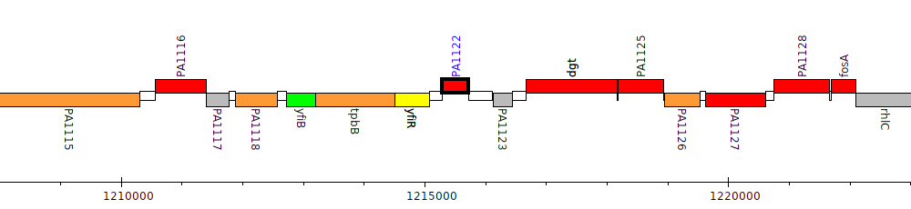
Gene Feature Overview
| Strain |
Pseudomonas aeruginosa PAO1 (Stover et al., 2000)
GCF_000006765.1|latest |
| Locus Tag |
PA1122
|
| Name |
Synonym: def Synonym: fms Synonym: pdf |
| Replicon | chromosome |
| Genomic location | 1215284 - 1215727 (+ strand) |
| Transposon Mutants | 3 transposon mutants in PAO1 |
| Transposon Mutants in orthologs | 1 transposon mutants in orthologs |
Cross-References
| RefSeq | NP_249813 |
| GI | 15596319 |
| Affymetrix | PA1122_at |
| Entrez | 878310 |
| GenBank | AAG04511.1 |
| NCBI Locus Tag | PA1122 |
| protein_id(GenBank) | gb|AAG04511.1|AE004542_7|gnl|PseudoCAP|PA1122 |
| TIGR | NTL03PA01123 |
| UniProtKB Acc | Q9I4L3 |
| UniProtKB ID | Q9I4L3_PSEAE |
Product
| Feature Type | CDS |
| Coding Frame | 1 |
| Product Name |
putative peptide deformylase
|
| Synonyms | |
| Evidence for Translation |
Identified using nanoflow high-pressure liquid chromatography (HPLC) in conjunction with microelectrospray ionization on LTQ XL mass spectrometer (PMID:24291602).
|
| Charge (pH 7) | -4.34 |
| Kyte-Doolittle Hydrophobicity Value | -0.307 |
| Molecular Weight (kDa) | 16.3 |
| Isoelectric Point (pI) | 4.99 |
Subcellular localization
| Individual Mappings | |
| Additional evidence for subcellular localization |
PDB 3D Structures
| Accession | Header | Accession Date | Compound | Source | Resolution | Method | Percent Identity |
| 6JFE | HYDROLASE | 02/08/19 | K2U bound crystal structure of class I type b peptide deformylase from Pseudomonas aeruginosa | Pseudomonas aeruginosa | 2.1 | X-RAY DIFFRACTION | 98.4 |
| 6JFC | HYDROLASE | 02/08/19 | Actinonin bound crystal structure of class I type b peptide deformylase from Pseudomonas aeruginosa | Pseudomonas aeruginosa | 2.04 | X-RAY DIFFRACTION | 98.4 |
| 6JFA | HYDROLASE | 02/08/19 | Met-Ala-Ser bound crystal structure of class I type b peptide deformylase from Pseudomonas aeruginosa | Pseudomonas aeruginosa; SYNTHETIC CONSTRUCT | 1.93 | X-RAY DIFFRACTION | 98.4 |
| 6JF9 | HYDROLASE | 02/08/19 | Apo crystal structure of class I type b peptide deformylase from Pseudomonas aeruginosa | Pseudomonas aeruginosa | 1.7 | X-RAY DIFFRACTION | 98.4 |
| 6JFD | HYDROLASE | 02/08/19 | K1U bound crystal structure of class I type b peptide deformylase from Pseudomonas aeruginosa | Pseudomonas aeruginosa | 2.4 | X-RAY DIFFRACTION | 98.4 |
| 6JFN | HYDROLASE | 02/11/19 | K4U bound crystal structure of class I type b peptide deformylase from Pseudomonas aeruginosa | Pseudomonas aeruginosa | 2.04 | X-RAY DIFFRACTION | 98.4 |
| 6JFF | HYDROLASE | 02/08/19 | K3U bound crystal structure of class I type b peptide deformylase from Pseudomonas aeruginosa | Pseudomonas aeruginosa | 2.1 | X-RAY DIFFRACTION | 98.4 |
Pathogen Association Analysis
| Results |
Common
Found in both pathogen and nonpathogenic strains
Hits to this gene were found in 568 genera
|
Orthologs/Comparative Genomics
| Pseudomonas Ortholog Database | View orthologs at Pseudomonas Ortholog Database |
| Pseudomonas Ortholog Group |
POG001082 (483 members) |
| Putative Inparalogs | None Found |
Interactions
| STRING database | Search for predicted protein-protein interactions using:
Search term: PA1122
Search term: putative peptide deformylase
|
Human Homologs
References
|
Evidence that peptide deformylase and methionyl-tRNA(fMet) formyltransferase are encoded within the same operon in Escherichia coli.
Meinnel T, Blanquet S
J Bacteriol 1993 Dec;175(23):7737-40
PubMed ID: 8244948
|
|
A new subclass of the zinc metalloproteases superfamily revealed by the solution structure of peptide deformylase.
Meinnel T, Blanquet S, Dardel F
J Mol Biol 1996 Sep 27;262(3):375-86
PubMed ID: 8845003
|